MAKE A MEME
View Large Image

| View Original: | HLA-B*5101.png (263x314) | |||
| Download: | Original | Medium | Small | Thumb |
| Courtesy of: | commons.wikimedia.org | More Like This | ||
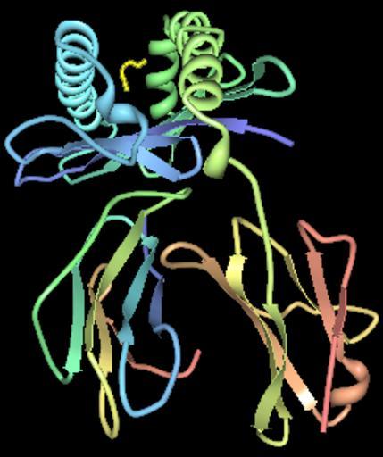
| Keywords: HLA-B*5101.png 1e28 Peptides is shown in yellow in the binding pocket Beta 2 microglobulin is show in the lower left and membrane attachment site is in the lower right 3D Structure is derived from Maenaka K et al 2000 Nonstandard peptide binding revealed by crystal structures of HLA-B 5101 complexed with HIV immunodominant epitopes J Immunol 165 3260-3267 own Pdeitiker HLA-B Cell surface antigens | ||||